Francis Y.H. Tien Vascular Centre

Non-invasive DIAGNOSTIC PROCEDURES
Doppler Evaluation
The Doppler effect refers to a frequency change when ultrasound is reflected from moving objects. By tracking the direction and velocity of red blood cells with this information, the Doppler probe can detect blood flow through vessels. Combined with sound spectral analysis and systolic blood pressure measurement, this technology is sensitive in identifying a narrowing, or stenosis, in an artery.
A comparison of the pressure taken at the ankle with that at the brachial artery is a good indication of whether any part of the lower limb arteries is significantly affected by atherosclerosis. When indicated of the disease, spectral waveforms and pressures taken segmentally would allow localization of the stenosis. The exercise test is excellent for revealing pressure reduction in patients with intermittent claudication. The cold stimulation test measures digital pressures following induced vasoconstriction for distinguishing Raynaud’s disease from upper limb arterial stenosis.
- Ankle-Brachial Index (ABI)
-
Segmental Waveforms and Pressures
Indications:
- Claudication
- Rest pain
- Ulcer or gangrene
- Treadmill Exercise Test
- Digital Artery Evaluation
-
Cold Stimulation Test
Indications:
- Digital colour changes

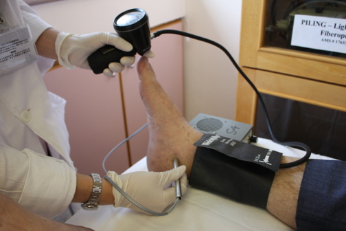
Measuring the ankle systolic pressure with a continuous-wave Doppler probe, pneumatic cuff and gauge. This reading would be compared with that at the brachial artery. Since pressure drops with increasing arterial stenosis, an ankle-brachial index of less than 0.95 would be considered abnormal.
Duplex Scan
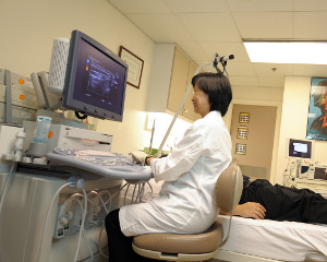
Duplex ultrasound is a combination of the Doppler technology with pulse-echo imaging. After sending off ultrasound pulses into the tissue, the instrument converts the echoes received from various locations into dots of varying strength, forming an image of the blood vessel.
Since duplex scanning can offer localization as well as physiological information of blood vessels, it is valuable for pre-operative evaluations. For example, it is an accurate while non-invasive method in revealing significant stenosis in the cervical carotid arteries, in which case operation may be needed. For those patients with abdominal aortic aneurysms, the state of the dilatation is followed up biannually until the need for surgery is deemed necessary. This technology is also beneficial to post-operative individuals whose graft or stent patency can be monitored on an out-patient basis.
-
Carotid and Vertebral Scan
Indications:
- Stroke
- Transient ischaemic attack
- High risk screening
-
Abdominal Scan
Indications:
- Severe hypertension
- Abdominal aortic aneurysm
- Graft Surveillance
- Stent Surveillance
- Detection for Deep Vein Thrombosis
Indications:
- lower limb oedema / pain
- Evaluation of Venous Valve Competence
Indications:
- Varicose vein
- Venous ulcer

Duplex scanning is a painless and cost-effective procedure. Image is obtained by putting an ultrasound probe onto the skin surface.

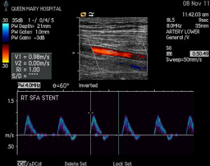
Duplex image of a superficial femoral artery post angioplasty and insertion of a nitinol stent.
Air-Plethysmography (APG)
- Detection for Chronic Venous Obstruction
-
Measurement of Venous Reflux
Indications:
- Varicose veins
- Venous ulcer
The APG quantifies the volume change in a limb caused by alterations in blood pressure. This information would indicate the presence of obstruction by venous thrombosis and the degree of venous reflux in the lower limb as a result of chronic venous insufficiency.

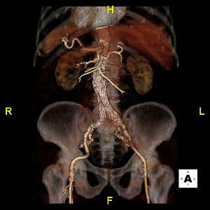
CT angiography with 3D reconstruction of abdominal aortic aneurysm post- endovascular repair.


